Our groundbreaking online platform offers a virtual anatomical dissection simulation and in-depth anatomical studies for students and educators alike. We have pioneered specialty scanners capable of capturing high-resolution photographic images of meticulously dissected specimens in a unique spherical mode. These images are then reconstructed into interactive, multi-layered, multidimensional sequences, which are made accessible through our dedicated website.
Unlike traditional animated 3D anatomy computer models, 4D Interactive Anatomy provides an unparalleled real dissection experience that can be reproduced anytime, anywhere. Students and users have the ability to browse these scans, select specimens, and engage in an immersive dissection process. With the ability to tilt, rotate, and peel away layer-by-layer to identify anatomical structures, our technology offers an authentic and hands-on learning experience like no other.
Our technology is based on a few critical assets:
Our partnerships with institutions
The donors
Our purpose-built scanners that capture the high-resolution images
Our colleagues labeling the images
Our partners handling translations & other content co-operations
Scanning Partnerships with Institutions
The advantages of incorporating 4D Anatomy Scanners into dissection labs are numerous. You can harness the power of this innovative technology to:
Create your own dissection scans and educational tools, empowering educators to develop customized teaching materials that align with your curriculum.
Archive and digitize specimens from anatomical museums, preserving these invaluable resources for future generations while making them accessible to a wider audience.
Utilize 4D Anatomy technology for the creation of publications and PhD works, enabling researchers to showcase their findings in an interactive and visually stunning format.
Explore anatomical variations, pathologies, and surgical techniques through dissection and scanning, offering your students the opportunity to delve deeper into specialized areas of study.
Facilitate dissection courses where your students are assigned a comprehensive dissection plan, using our scanners to document their own dissections and create state-of-the-art interactive educational material for your institution.
Engage your students in laboratory work like never before, igniting their passion for anatomical exploration and fostering a deeper understanding of the human body.
By starting a scanning partnership with us, you can join an elite society where dissections are showcased to the most prestigious universities worldwide through the www.4danatomy.com website. Experience the future of anatomical education with 4D Interactive Anatomy and empower your students to excel in their studies like never before.
Use Case from the University of Debrecen
We are currently partnered with the University of Debrecen, Faculty of Medicine, Department of Anatomy, Histology, and Embryology for scanning new content.
Dr. Tamás Juhász and Dr. Péter Szűcs organized an elective “4D Anatomy” dissection course that a small group of students may take each semester. The students receive specimens and dissection plans they need to follow under the faculty's guidance.
Students are eager to join the course as they can develop their skills and record their work with our scanner for longevity. If their work is up to the standards of 4D Interactive Anatomy, we publish the scan on our website, crediting the students, faculty, and University as the authors.
As Dániel Kovács, one of the students participating in the Fall 2022 4D Anatomy course who created the “Brain (lateral ventricle)” scan published on our website said:
"Thanks to the state-of-the-art tools and professional advice during the 4D Anatomy course, we were able to further enhance our understanding of human anatomy that we previously developed during the lab work. Dr. Tamás Juhász guided us throughout the course and was always happy to answer our questions. I improved my precision, fine motor- and technical skills in a fun and engaging way. I was motivated to have my work published on the website. Now, I can use it to study at any time.”
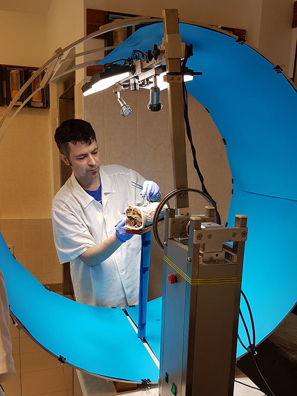
Scanning Process in Practice
Students scan the specimens in stages (layer-by-layer) throughout the dissection process. The first step is to secure the specimen to the rotating table and place it into the scanner. Next is starting the scanning process.
It takes about 20 minutes for the scanner to scan the first layer of the specimen. It takes another ~30 minutes for the cloud processing to complete processing. Once the cloud processing is done, you can access the scan through our administrator website. The scan is not yet published to the general user base at this stage.
Once done the scanning is done (even if the cloud processing is not completed), the specimen may be removed from the scanner and the next stage of dissection is performed. The specimen is then placed back into the scanner to start the scanning of the next layer. The steps of scanning and dissecting are repeated until the specimen is dissected down to the last layer.
Multiple students can work on multiple specimens at the same time, as the scanner itself is only actually in use for the period of running the scan (~20 minutes).
Once all layers of a specimen are scanned, we adjust the images as needed and start labeling the anatomical structures on the scan.
Interested in Having One of Our Scanners at Your Institution?
What our customers are saying
“It is amazing to see how engaged students are when doing their dissections and scanning in the 4D Anatomy elective course.”
Tamás Juhász PhD
Adjunct
University of Debrecen

“I improved my precision, fine motor- and technical skills in a fun and engaging way. I was motivated to have my work published on the website. Now, I can use it to study at any time”
Dániel Kovács
Student, 4D Anatomy Course participant
Donor Acknowledgement
We place our scanners at institutions that have a willed body donor program to ensure that we are allowed to record the images for educational & research purposes in line with the donors' original will. For more information about our partner institutions' donor programs, please visit their websites:
Semmelweis University: https://semmelweis.hu/anatomia/tetemfelajanlas-aktualis/
University of Debrecen: https://anat.unideb.hu/hu/akik-holtukban-az-eloket-szolgaljak
University of Szeged: https://www.med.u-szeged.hu/anatomy/holttest-felajanlas/holttest-felajanlas
The donor statements and related records are kept at and by the institutions.

Donor acknowledgement:
4D Interactive Anatomy is grateful to the donors who make this website possible. Their altruistic sacrifice helped tens of thousands of students so far to get a better anatomy education. Please always take care to respect the donors at all times when accessing the website and using the images & contents.
The images of donors may be disturbing to some viewers. Please take appropriate measures at all times to protect the donors dignity and privacy. When using the website you agree to adhere to our Terms of Use. If you have any questions about the donors and use of images, please contact us.
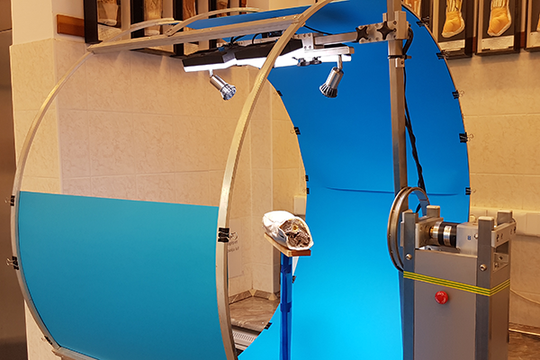
Our Scanners
To capture the amazing details you can see in our scans, we are using photography-based scanning. Our scanners have two axis of rotation that allows us to capture images at different angles of rotation. We typically create half-sphere scans and then flip over the specimen to scan the other side or use a different specimen for dissecting from the other side.
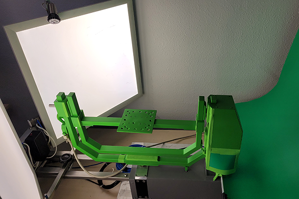
NeuroArc V1
Our first fully motorized scanner “NeuroArc V1” was used to capture many of the highly detailed scans you can still find on our website, such as the head & neck with upper chest and head & neck with back scans.
This scanner can scan up to 30 x 40 cm specimens. Due to the way it was constructed, the specimen is rotated to its side as the camera is in a fixed position. Therefore, stably fixating the specimen is necessary.
Currently located at the University of Szeged in Hungary, this scanner is being used for PhD research and work related to endoscopic neurosurgical techniques simulated on donors.
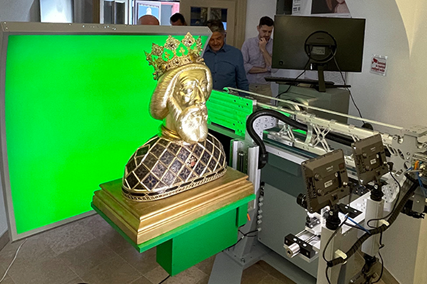
NeuroArc V2
Our second generation, fully motorized scanner “NeuroArc V2” was created to scan larger specimens. Compared to the V1 scanner, this one is both larger and heavier.
Unlike the V1 scanner, this has the camera on a rotating arm, so the specimen is not turned to its side while scanning. This allows for scanning wet specimens and other delicate prosections.
This scanner was extensively used at the University of Debrecen to scan many of the bones and some dissections you can find on our website.
It is currently being upgraded and is not deployed at an institution.
NeuroArc V3
Our newest scanner, the “NeuroArc V3” is a precision machine. Capable of scanning even larger specimens, this is a state-of-the-art device.
Currently located at the University of Debrecen, this scanner is used for continuos content production. We are also using it to digitize the anatomical (museum) collection of the University.
The largest and heaviest of our scanners, this is also the most precise, allowing for fine-tuning settings for scanning.
As this scanner comes with a production plan, we can manufacture more of them to keep adding new scanners into our content production network.
Labelling the Images
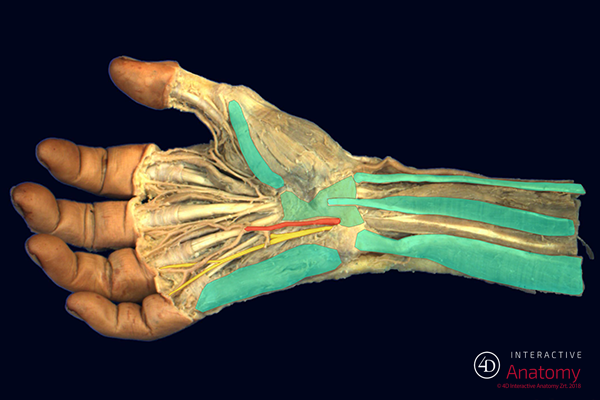
45000+ labels by professionals
There are over 45000 labels denoting anatomical structures on our images. These labels are hand-drawn over the images by our professional medical staff and partners:
- Tamás Juhász PhD., Adjunct, Department of Anatomy and Cell Biology at the University of Debrecen
- Eszter Orbán, Midwife, Specialist at 4D Interactive Anatomy
- Dr. Attila Álmos Balogh PhD., Neurosurgeon, CEO at 4D Interactive Anatomy
- Dr. Máté Borbély, Specialist at 4D Interactive Anatomy
The labeling is peer-reviewed to ensure the highest standards and accuracy. The labels are sorted into one of the 18 categories, each marked with a different color. Once a scan is labeled, it is released to the general user base of 4D Interactive Anatomy.
Translations
3500+ structures in 4 languages
The English, Latin, Spanish, and Ukrainian language translations of the 3500+ anatomical structures on our website are maintained by our friends and colleagues. We thank them for their endless efforts:
Professor D.Med.S. Oleksandr Kovalchuk, and colleagues of the Department of Anatomy and Pathological Physiology at Educational And Scientific Center "Institute of Biology and Medicine" of Taras Shevchenko National University of Kyiv
Tamás Juhász PhD., Adjunct, Department of Anatomy and Cell Biology at the University of Debrecen
Dr. Attila Álmos Balogh PhD., Neurosurgeon, CEO at 4D Interactive Anatomy
Dr. Máté Borbély, Specialist at 4D Interactive Anatomy
We are always looking for partnerships. If you are an expert in anatomical terminology in your own language and would be interested in getting access to 4D Interactive Anatomy in exchange for translating our structures database, please contact us.